Background
Appendicitis is defined as an inflammation of the inner lining of the vermiform appendix that spreads to its other parts. This condition is a common and urgent surgical illness with protean manifestations, generous overlap with other clinical syndromes, and significant morbidity, which increases with diagnostic delay (see Clinical Presentation). In fact, despite diagnostic and therapeutic advancement in medicine, appendicitis remains a clinical emergency and is one of the more common causes of acute abdominal pain.
No single sign, symptom, or diagnostic test accurately confirms the diagnosis of appendiceal inflammation in all cases, and the classic history of anorexia and periumbilical pain followed by nausea, right lower quadrant (RLQ) pain, and vomiting occurs in only 50% of cases (see Clinical Presentation).
Appendicitis may occur for several reasons, such as an infection of the appendix, but the most important factor is the obstruction of the appendiceal lumen (see Pathogenesis and Etiology). Left untreated, appendicitis has the potential for severe complications, including perforation or sepsis, and may even cause death (see Prognosis and Complications). However, the differential diagnosis of appendicitis is often a clinical challenge because appendicitis can mimic several abdominal conditions (see Diagnostic Considerations and Differentials).[1]
Appendectomy remains the only curative treatment of appendicitis (see Treatment and Management). The surgeon's goals are to evaluate a relatively small population of patients referred for suspected appendicitis and to minimize the negative appendectomy rate without increasing the incidence of perforation. The emergency department (ED) clinician must evaluate the larger group of patients who present to the ED with abdominal pain of all etiologies with the goal of approaching 100% sensitivity for the diagnosis in a time-, cost-, and consultation-efficient manner.
Go to Pediatric Appendicitis for more information on this topic.
Anatomy
The appendix is a wormlike extension of the cecum and, for this reason, has been called the vermiform appendix. The average length of the appendix is 8-10 cm (ranging from 2-20 cm). The appendix appears during the fifth month of gestation, and several lymphoid follicles are scattered in its mucosa. Such follicles increase in number when individuals are aged 8-20 years. A normal appendix is seen below.
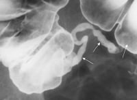 Normal appendix; barium enema radiographic examination. A complete contrast-filled appendix is observed (arrows), which effectively excludes the diagnosis of appendicitis. The appendix is contained within the visceral peritoneum that forms the serosa, and its exterior layer is longitudinal and derived from the taenia coli; the deeper, interior muscle layer is circular. Beneath these layers lies the submucosal layer, which contains lymphoepithelial tissue. The mucosa consists of columnar epithelium with few glandular elements and neuroendocrine argentaffin cells.
Taenia coli converge on the posteromedial area of the cecum, which is the site of the appendiceal base. The appendix runs into a serosal sheet of the peritoneum called the mesoappendix, within which courses the appendicular artery, which is derived from the ileocolic artery. Sometimes, an accessory appendicular artery (deriving from the posterior cecal artery) may be found.
Normal appendix; barium enema radiographic examination. A complete contrast-filled appendix is observed (arrows), which effectively excludes the diagnosis of appendicitis. The appendix is contained within the visceral peritoneum that forms the serosa, and its exterior layer is longitudinal and derived from the taenia coli; the deeper, interior muscle layer is circular. Beneath these layers lies the submucosal layer, which contains lymphoepithelial tissue. The mucosa consists of columnar epithelium with few glandular elements and neuroendocrine argentaffin cells. Taenia coli converge on the posteromedial area of the cecum, which is the site of the appendiceal base. The appendix runs into a serosal sheet of the peritoneum called the mesoappendix, within which courses the appendicular artery, which is derived from the ileocolic artery. Sometimes, an accessory appendicular artery (deriving from the posterior cecal artery) may be found.
Appendiceal vasculature
The vasculature of the appendix must be addressed to avoid intraoperative hemorrhages. The appendicular artery is contained within the mesenteric fold that arises from a peritoneal extension from the terminal ileum to the medial aspect of the cecum and appendix; it is a terminal branch of the ileocolic artery and runs adjacent to the appendicular wall. Venous drainage is via the ileocolic veins and the right colic vein into the portal vein; lymphatic drainage occurs via the ileocolic nodes along the course of the superior mesenteric artery to the celiac nodes and cisterna chyli.Appendiceal location
The appendix has no fixed position. It originates 1.7-2.5 cm below the terminal ileum, either in a dorsomedial location (most common) from the cecal fundus, directly beside the ileal orifice, or as a funnel-shaped opening (2-3% of patients). The appendix has a retroperitoneal location in 65% of patients and may descend into the iliac fossa in 31%. In fact, many individuals may have an appendix located in the retroperitoneal space; in the pelvis; or behind the terminal ileum, cecum, ascending colon, or liver. Thus, the course of the appendix, the position of its tip, and the difference in appendiceal position considerably changes clinical findings, accounting for the nonspecific signs and symptoms of appendicitis.Congenital appendiceal disorders
Appendiceal congenital disorders are extremely rare but occasionally reported (eg, agenesis, duplication, triplication).Pathophysiology
Reportedly, appendicitis is caused by obstruction of the appendiceal lumen from a variety of causes (see Etiology). Independent of the etiology, obstruction is believed to cause an increase in pressure within the lumen. Such an increase is related to continuous secretion of fluids and mucus from the mucosa and the stagnation of this material. At the same time, intestinal bacteria within the appendix multiply, leading to the recruitment of white blood cells (see the image below) and the formation of pus and subsequent higher intraluminal pressure.
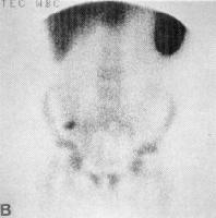 Technetium-99m radionuclide scan of the abdomen shows focal uptake of labeled WBCs in the right lower quadrant consistent with acute appendicitis. If appendiceal obstruction persists, intraluminal pressure rises ultimately above that of the appendiceal veins, leading to venous outflow obstruction. As a consequence, appendiceal wall ischemia begins, resulting in a loss of epithelial integrity and allowing bacterial invasion of the appendiceal wall.
Within a few hours, this localized condition may worsen because of thrombosis of the appendicular artery and veins, leading to perforation and gangrene of the appendix. As this process continues, a periappendicular abscess or peritonitis may occur.
Technetium-99m radionuclide scan of the abdomen shows focal uptake of labeled WBCs in the right lower quadrant consistent with acute appendicitis. If appendiceal obstruction persists, intraluminal pressure rises ultimately above that of the appendiceal veins, leading to venous outflow obstruction. As a consequence, appendiceal wall ischemia begins, resulting in a loss of epithelial integrity and allowing bacterial invasion of the appendiceal wall. Within a few hours, this localized condition may worsen because of thrombosis of the appendicular artery and veins, leading to perforation and gangrene of the appendix. As this process continues, a periappendicular abscess or peritonitis may occur.
Etiology
Appendicitis is caused by obstruction of the appendiceal lumen. The most common causes of luminal obstruction include lymphoid hyperplasia secondary to inflammatory bowel disease (IBD) or infections (more common during childhood and in young adults), fecal stasis and fecaliths (more common in elderly patients), parasites (especially in Eastern countries), or, more rarely, foreign bodies and neoplasms.
Fecaliths form when calcium salts and fecal debris become layered around a nidus of inspissated fecal material located within the appendix. Lymphoid hyperplasia is associated with various inflammatory and infectious disorders including Crohn disease, gastroenteritis, amebiasis, respiratory infections, measles, and mononucleosis.
Obstruction of the appendiceal lumen has less commonly been associated with bacteria (Yersinia species, adenovirus, cytomegalovirus, actinomycosis, Mycobacteria species, Histoplasma species), parasites (eg, Schistosomes species, pinworms, Strongyloides stercoralis), foreign material (eg, shotgun pellet, intrauterine device, tongue stud, activated charcoal), tuberculosis, and tumors.
Epidemiology
Appendicitis is one of the more common surgical emergencies, and it is one of the most common causes of abdominal pain. In the United States, 250,000 cases of appendicitis are reported annually, representing 1 million patient-days of admission. The incidence of acute appendicitis has been declining steadily since the late 1940s, and the current annual incidence is 10 cases per 100,000 population. Appendicitis occurs in 7% of the US population, with an incidence of 1.1 cases per 1000 people per year. Some familial predisposition exists.
In Asian and African countries, the incidence of acute appendicitis is probably lower because of the dietary habits of the inhabitants of these geographic areas. The incidence of appendicitis is lower in cultures with a higher intake of dietary fiber. Dietary fiber is thought to decrease the viscosity of feces, decrease bowel transit time, and discourage formation of fecaliths, which predispose individuals to obstructions of the appendiceal lumen.
In the last few years, a decrease in frequency of appendicitis in Western countries has been reported, which may be related to changes in dietary fiber intake. In fact, the higher incidence of appendicitis is believed to be related to poor fiber intake in such countries.
There is a slight male preponderance of 3:2 in teenagers and young adults; in adults, the incidence of appendicitis is approximately 1.4 times greater in men than in women. The incidence of primary appendectomy is approximately equal in both sexes.
The incidence of appendicitis gradually rises from birth, peaks in the late teen years, and gradually declines in the geriatric years. The mean age when appendicitis occurs in the pediatric population is 6-10 years. Lymphoid hyperplasia is observed more often among infants and adults and is responsible for the increased incidence of appendicitis in these age groups. Younger children have a higher rate of perforation, with reported rates of 50-85%. The median age at appendectomy is 22 years. Although rare, neonatal and even prenatal appendicitis have been reported. Clinicians must maintain a high index of suspicion in all age groups.
Go to Pediatric Appendicitis for more information on this topic.
Prognosis
Acute appendicitis is the most common reason for emergency abdominal surgery. Appendectomy carries a complication rate of 4-15%, as well as associated costs and the discomfort of hospitalization and surgery. Therefore, the goal of the surgeon is to make an accurate diagnosis as early as possible. Delayed diagnosis and treatment account for much of the mortality and morbidity associated with appendicitis.
The overall mortality rate of 0.2-0.8% is attributable to complications of the disease rather than to surgical intervention. The mortality rate in children ranges from 0.1% to 1%; in patients older than 70 years, the rate rises above 20%, primarily because of diagnostic and therapeutic delay.
Appendiceal perforation is associated with increased morbidity and mortality compared with nonperforating appendicitis. The mortality risk of acute but not gangrenous appendicitis is less than 0.1%, but the risk rises to 0.6% in gangrenous appendicitis. The rate of perforation varies from 16% to 40%, with a higher frequency occurring in younger age groups (40-57%) and in patients older than 50 years (55-70%), in whom misdiagnosis and delayed diagnosis are common. Complications occur in 1-5% of patients with appendicitis, and postoperative wound infections account for almost one third of the associated morbidity.
Other problems that should be considered in a patient with suspected appendicitis include appendiceal stump appendicitis, typhlitis, epiploic appendagitis, psoas abscess, and yersiniosis.
Misdiagnosis in women of childbearing age
Appendicitis is misdiagnosed in 33% of nonpregnant women of childbearing age. The most frequent misdiagnoses are PID, followed by gastroenteritis and urinary tract infection. In distinguishing appendiceal pain from that of PID, anorexia and onset of pain more than 14 days after menses suggests appendicitis. Previous PID, vaginal discharge, or urinary symptoms indicates PID. On physical examination, tenderness outside the RLQ, cervical motion tenderness, vaginal discharge, and positive urinalysis support the diagnosis of PID.
Although negative appendectomy does not appear to adversely affect maternal or fetal health, diagnostic delay with perforation does increase fetal and maternal morbidity. Therefore, aggressive evaluation of the appendix is warranted in pregnant women.
The level of urinary beta–human chorionic gonadotropin (beta-hCG) is useful in differentiating appendicitis from early ectopic pregnancy. However, with regard to the WBC count, physiologic leukocytosis during pregnancy makes this study less useful in the diagnosis than at other times, and no reliable distinguishing WBC parameters are cited in the literature.
Misdiagnosis in children
Appendicitis is misdiagnosed in 25-30% of children, and the rate of initial misdiagnosis is inversely related to the age of the patient. The most common misdiagnosis is gastroenteritis, followed by upper respiratory infection and lower respiratory infection.
Children with misdiagnosed appendicitis are more likely than their counterparts to have vomiting before pain onset, diarrhea, constipation, dysuria, signs and symptoms of upper respiratory infection, and lethargy or irritability. Physical findings less likely to be documented in children with a misdiagnosis than in others include bowel sounds; peritoneal signs; rectal findings; and ear, nose, and throat findings.
Considerations in elderly patients
Appendicitis in patients older than 60 years accounts for 10% of all appendectomies. The incidence of misdiagnosis is increased in elderly patients.
Older patients tend to seek medical attention later in the course of illness; therefore, a duration of symptoms in excess of 24-48 hours should not dissuade the clinician from the diagnosis. In patients with comorbid conditions, diagnostic delay is correlated with increased morbidity and mortality.
The overall mortality rate of 0.2-0.8% is attributable to complications of the disease rather than to surgical intervention. The mortality rate in children ranges from 0.1% to 1%; in patients older than 70 years, the rate rises above 20%, primarily because of diagnostic and therapeutic delay.
Appendiceal perforation is associated with increased morbidity and mortality compared with nonperforating appendicitis. The mortality risk of acute but not gangrenous appendicitis is less than 0.1%, but the risk rises to 0.6% in gangrenous appendicitis. The rate of perforation varies from 16% to 40%, with a higher frequency occurring in younger age groups (40-57%) and in patients older than 50 years (55-70%), in whom misdiagnosis and delayed diagnosis are common. Complications occur in 1-5% of patients with appendicitis, and postoperative wound infections account for almost one third of the associated morbidity.
History
Variations in the position of the appendix, age of the patient, and degree of inflammation make the clinical presentation of appendicitis notoriously inconsistent. Statistics report that 1 of 5 cases of appendicitis is misdiagnosed; however, a normal appendix is found in 15-40% of patients who have an emergency appendectomy.
Niwa et al reported an interesting case of a young woman with recurrent pain in who was referred for appendicitis, treated with antibiotics, and was found to have an appendiceal diverticulitis associated with a rare pelvic pseudocyst at laparotomy after 12 months.[2] Her condition was probably due to diverticular perforation of the pseudocyst.
Symptoms
The classic history of anorexia and periumbilical pain followed by nausea, right lower quadrant (RLQ) pain, and vomiting occurs in only 50% of cases. Nausea is present in 61-92% of patients; anorexia is present in 74-78% of patients. Neither finding is statistically different from findings in patients who present to the emergency department with other etiologies of abdominal pain. In addition, when vomiting occurs, it nearly always follows the onset of pain. Vomiting that precedes pain is suggestive of intestinal obstruction, and the diagnosis of appendicitis should be reconsidered. Diarrhea or constipation is noted in as many as 18% of patients and should not be used to discard the possibility of appendicitis.
The most common symptom of appendicitis is abdominal pain. Typically, symptoms begin as periumbilical or epigastric pain migrating to the right lower quadrant (RLQ) of the abdomen. This pain migration is the most discriminating feature of the patient's history, with a sensitivity and specificity of approximately 80%, a positive likelihood ratio of 3.18, and a negative likelihood ratio of 0.5.[3] Patients usually lie down, flex their hips, and draw their knees up to reduce movements and to avoid worsening their pain. Later, a worsening progressive pain along with vomiting, nausea, and anorexia are described by the patient. Usually, a fever is not present at this stage.
The duration of symptoms is less than 48 hours in approximately 80% of adults but tends to be longer in elderly persons and in those with perforation. Approximately 2% of patients report duration of pain in excess of 2 weeks. A history of similar pain is reported in as many as 23% of cases, but this history of similar pain, in and of itself, should not be used to rule out the possibility of appendicitis.
In addition to recording the history of the abdominal pain, obtain a complete summary of the recent personal history surrounding gastroenterologic, genitourinary, and pneumologic conditions, as well as consider gynecologic history in female patients. An inflamed appendix near the urinary bladder or ureter can cause irritative voiding symptoms and hematuria or pyuria. Cystitis in male patients is rare in the absence of instrumentation. Consider the possibility of an inflamed pelvic appendix in male patients with apparent cystitis. Also consider the possibility of appendicitis in pediatric or adult patients who present with acute urinary retention.[4] Physical Examination
It is important to remember that the position of the appendix is variable. Of 100 patients undergoing 3-dimensional (3-D) multidetector computed tomography (MDCT) scanning, the base of the appendix was located at the McBurney point in only 4% of patients; in 36%, the base was within 3 cm of the point; in 28%, it was 3-5 cm from that point; and, in 36% of patients, the base of the appendix was more than 5 cm from the McBurney point.[5]
The most specific physical findings in appendicitis are rebound tenderness, pain on percussion, rigidity, and guarding. Although RLQ tenderness is present in 96% of patients, this is a nonspecific finding. Rarely, left lower quadrant (LLQ) tenderness has been the major manifestation in patients with situs inversus or in patients with a lengthy appendix that extends into the LLQ. Tenderness on palpation in the RLQ over the McBurney point is the most important sign in these patients.
A careful physical examination, not limited to the abdomen, must be performed in any patient with suspected appendicitis. Gastrointestinal (GI), genitourinary, and pulmonary systems must be studied. Male infants and children occasionally present with an inflamed hemiscrotum due to migration of an inflamed appendix or pus through a patent processus vaginalis. This is often initially misdiagnosed as acute testicular torsion. In addition, perform a rectal examination in any patient with an unclear clinical picture, and perform a pelvic examination in all women with abdominal pain.
According to the American College of Emergency Physicians (ACEP) 2010 clinical policy update, clinical signs and symptoms should be used to stratify patient risk and to choose next steps for testing and management.[6, 7]
Accessory signs
In a minority of patients with acute appendicitis, some other signs may be noted. However, their absence never should be used to rule out appendiceal inflammation. The Rovsing sign (RLQ pain with palpation of the LLQ) suggests peritoneal irritation in the RLQ precipitated by palpation at a remote location. The obturator sign (RLQ pain with internal and external rotation of the flexed right hip) suggests that the inflamed appendix is located deep in the right hemipelvis. The psoas sign (RLQ pain with extension of the right hip or with flexion of the right hip against resistance) suggests that an inflamed appendix is located along the course of the right psoas muscle.
The Dunphy sign (sharp pain in the RLQ elicited by a voluntary cough) may be helpful in making the clinical diagnosis of localized peritonitis. Similarly, RLQ pain in response to percussion of a remote quadrant of the abdomen, or to firm percussion of the patient's heel, suggests peritoneal inflammation.
The Markle sign, pain elicited in a certain area of the abdomen when the standing patient drops from standing on toes to the heels with a jarring landing, was studied in 190 patients undergoing appendectomy and found to have a sensitivity of 74%.[8]
Rectal examination
There is no evidence in the medical literature that the digital rectal examination (DRE) provides useful information in the evaluation of patients with suspected appendicitis; however, failure to perform a rectal examination is frequently cited in successful malpractice claims. In 2008, Sedlak et al studied 577 patients who underwent DRE as part of an evaluation for suspected appendicitis and found no value as a means of distinguishing patients with and without appendicitis.[9] Appendicitis and Pregnancy
The incidence of appendicitis is unchanged in pregnancy relative to the general population, but the clinical presentation is more variable than at other times.
During pregnancy, the appendix migrates in a counterclockwise direction toward the right kidney, rising above the iliac crest at about 4.5 months' gestation. RLQ pain and tenderness dominate in the first trimester, but in the latter half of pregnancy, right upper quadrant (RUQ) or right flank pain must be considered a possible sign of appendiceal inflammation.
Nausea, vomiting, and anorexia are common in uncomplicated first trimester pregnancies, but their reappearance later in gestation should be viewed with suspicion. Diagnostic Scoring
Several investigators have created diagnostic scoring systems to predict the likelihood of acute appendicitis. In these systems, a finite number of clinical variables is elicited from the patient and each is given a numeric value; then, the sum of these values is used.
The best known of these scoring systems is the MANTRELS score, which tabulates migration of pain, anorexia, nausea and/or vomiting, tenderness in the RLQ, rebound tenderness, elevated temperature, leukocytosis, and shift to the left (see Table 1).[10]
Table 1. MANTRELS Score (Open Table in a new window)
Characteristic Score M = Migration of pain to the RLQ 1 A = Anorexia 1 N = Nausea and vomiting 1 T = Tenderness in RLQ 2 R = Rebound pain 1 E = Elevated temperature 1 L = Leukocytosis 2 S = Shift of WBCs to the left 1 Total 10 Source: Alvarado.[10] RLQ = right lower quadrant; WBCs = white blood cells
Clinical scoring systems are attractive because of their simplicity; however, none has been shown prospectively to improve on the clinician's judgment in the subset of patients evaluated in the emergency department (ED) for abdominal pain suggestive of appendicitis. The MANTRELS score, in fact, was based on a population of patients hospitalized for suspected appendicitis, which differs markedly from the population seen in the ED.
In reviewing the records of 150 ED patients who underwent abdominopelvic computed tomography (CT) scanning to rule out appendicitis, McKay and Shepherd suggested that patients with an MANTRELS score of 0-3 could be discharged without imaging, that those with scores of 7 or above receive surgical consultation, and those with scores of 4-6 undergo CT evaluation.[11] The investigators found that patients with a MANTRELS score of 3 or lower had a 3.6% incidence of appendicitis, patients with scores of 4-6 had a 32% incidence of appendicitis, and patients with scores of 7-10 had a 78% incidence of appendicitis.[11]
In another study, Schneider et al concluded that the MANTRELS score was not sufficiently accurate to be used as the sole method for determining the need for appendectomy in the pediatric population.[12] These investigators, studied 588 patients aged 3-21 years and found that a MANTRELS score of 7 or greater had a positive predictive value of 65% and a negative predictive value of 85%.
Scoring systems and computer-aided diagnosis
Computer-aided diagnosis consists of using retrospective data of clinical features of patients with appendicitis and other causes of abdominal pain and then prospectively assessing the risk of appendicitis. Computer-aided diagnosis can achieve a sensitivity greater than 90% while reducing rates of perforation and negative laparotomy by as much as 50%.
However, the principle disadvantages to this method are that each institution must generate its own database to reflect characteristics of its local population, and specialized equipment and significant initiation time are required. In addition, computer-aided diagnosis is not widely available in US EDs. Stages of Appendicitis
The stages of appendicitis can be divided into early, suppurative, gangrenous, perforated, phlegmonous, spontaneous resolving, recurrent, and chronic.
Early stage appendicitis
In the early stage of appendicitis, obstruction of the appendiceal lumen leads to mucosal edema, mucosal ulceration, bacterial diapedesis, appendiceal distention due to accumulated fluid, and increasing intraluminal pressure. The visceral afferent nerve fibers are stimulated, and the patient perceives mild visceral periumbilical or epigastric pain, which usually lasts 4-6 hours.
Suppurative appendicitis
Increasing intraluminal pressures eventually exceed capillary perfusion pressure, which is associated with obstructed lymphatic and venous drainage and allows bacterial and inflammatory fluid invasion of the tense appendiceal wall. Transmural spread of bacteria causes acute suppurative appendicitis. When the inflamed serosa of the appendix comes in contact with the parietal peritoneum, patients typically experience the classic shift of pain from the periumbilicus to the right lower abdominal quadrant (RLQ), which is continuous and more severe than the early visceral pain.
Gangrenous appendicitis
Intramural venous and arterial thromboses ensue, resulting in gangrenous appendicitis.
Perforated appendicitis
Persisting tissue ischemia results in appendiceal infarction and perforation. Perforation can cause localized or generalized peritonitis.
Phlegmonous appendicitis or abscess
An inflamed or perforated appendix can be walled off by the adjacent greater omentum or small-bowel loops, resulting in phlegmonous appendicitis or focal abscess.
Spontaneously resolving appendicitis
If the obstruction of the appendiceal lumen is relieved, acute appendicitis may resolve spontaneously.[13, 14] This occurs if the cause of the symptoms is lymphoid hyperplasia or when a fecalith is expelled from the lumen.
Recurrent appendicitis
The incidence of recurrent appendicitis is 10%. The diagnosis is accepted as such if the patient underwent similar occurrences of RLQ pain at different times that, after appendectomy, were histopathologically proven to be the result of an inflamed appendix.
Chronic appendicitis
Chronic appendicitis occurs with an incidence of 1% and is defined by the following: (1) the patient has a history of RLQ pain of at least 3 weeks’ duration without an alternative diagnosis; (2) after appendectomy, the patient experiences complete relief of symptoms; (3) histopathologically, the symptoms were proven to be the result of chronic active inflammation of the appendiceal wall or fibrosis of the appendix.
History
Variations in the position of the appendix, age of the patient, and degree of inflammation make the clinical presentation of appendicitis notoriously inconsistent. Statistics report that 1 of 5 cases of appendicitis is misdiagnosed; however, a normal appendix is found in 15-40% of patients who have an emergency appendectomy.
Niwa et al reported an interesting case of a young woman with recurrent pain in who was referred for appendicitis, treated with antibiotics, and was found to have an appendiceal diverticulitis associated with a rare pelvic pseudocyst at laparotomy after 12 months.[2] Her condition was probably due to diverticular perforation of the pseudocyst.
The most common symptom of appendicitis is abdominal pain. Typically, symptoms begin as periumbilical or epigastric pain migrating to the right lower quadrant (RLQ) of the abdomen. This pain migration is the most discriminating feature of the patient's history, with a sensitivity and specificity of approximately 80%, a positive likelihood ratio of 3.18, and a negative likelihood ratio of 0.5.[3] Patients usually lie down, flex their hips, and draw their knees up to reduce movements and to avoid worsening their pain. Later, a worsening progressive pain along with vomiting, nausea, and anorexia are described by the patient. Usually, a fever is not present at this stage.
The duration of symptoms is less than 48 hours in approximately 80% of adults but tends to be longer in elderly persons and in those with perforation. Approximately 2% of patients report duration of pain in excess of 2 weeks. A history of similar pain is reported in as many as 23% of cases, but this history of similar pain, in and of itself, should not be used to rule out the possibility of appendicitis.
In addition to recording the history of the abdominal pain, obtain a complete summary of the recent personal history surrounding gastroenterologic, genitourinary, and pneumologic conditions, as well as consider gynecologic history in female patients. An inflamed appendix near the urinary bladder or ureter can cause irritative voiding symptoms and hematuria or pyuria. Cystitis in male patients is rare in the absence of instrumentation. Consider the possibility of an inflamed pelvic appendix in male patients with apparent cystitis. Also consider the possibility of appendicitis in pediatric or adult patients who present with acute urinary retention.[4]
Niwa et al reported an interesting case of a young woman with recurrent pain in who was referred for appendicitis, treated with antibiotics, and was found to have an appendiceal diverticulitis associated with a rare pelvic pseudocyst at laparotomy after 12 months.[2] Her condition was probably due to diverticular perforation of the pseudocyst.
Symptoms
The classic history of anorexia and periumbilical pain followed by nausea, right lower quadrant (RLQ) pain, and vomiting occurs in only 50% of cases. Nausea is present in 61-92% of patients; anorexia is present in 74-78% of patients. Neither finding is statistically different from findings in patients who present to the emergency department with other etiologies of abdominal pain. In addition, when vomiting occurs, it nearly always follows the onset of pain. Vomiting that precedes pain is suggestive of intestinal obstruction, and the diagnosis of appendicitis should be reconsidered. Diarrhea or constipation is noted in as many as 18% of patients and should not be used to discard the possibility of appendicitis.The most common symptom of appendicitis is abdominal pain. Typically, symptoms begin as periumbilical or epigastric pain migrating to the right lower quadrant (RLQ) of the abdomen. This pain migration is the most discriminating feature of the patient's history, with a sensitivity and specificity of approximately 80%, a positive likelihood ratio of 3.18, and a negative likelihood ratio of 0.5.[3] Patients usually lie down, flex their hips, and draw their knees up to reduce movements and to avoid worsening their pain. Later, a worsening progressive pain along with vomiting, nausea, and anorexia are described by the patient. Usually, a fever is not present at this stage.
The duration of symptoms is less than 48 hours in approximately 80% of adults but tends to be longer in elderly persons and in those with perforation. Approximately 2% of patients report duration of pain in excess of 2 weeks. A history of similar pain is reported in as many as 23% of cases, but this history of similar pain, in and of itself, should not be used to rule out the possibility of appendicitis.
In addition to recording the history of the abdominal pain, obtain a complete summary of the recent personal history surrounding gastroenterologic, genitourinary, and pneumologic conditions, as well as consider gynecologic history in female patients. An inflamed appendix near the urinary bladder or ureter can cause irritative voiding symptoms and hematuria or pyuria. Cystitis in male patients is rare in the absence of instrumentation. Consider the possibility of an inflamed pelvic appendix in male patients with apparent cystitis. Also consider the possibility of appendicitis in pediatric or adult patients who present with acute urinary retention.[4]
Physical Examination
It is important to remember that the position of the appendix is variable. Of 100 patients undergoing 3-dimensional (3-D) multidetector computed tomography (MDCT) scanning, the base of the appendix was located at the McBurney point in only 4% of patients; in 36%, the base was within 3 cm of the point; in 28%, it was 3-5 cm from that point; and, in 36% of patients, the base of the appendix was more than 5 cm from the McBurney point.[5]
The most specific physical findings in appendicitis are rebound tenderness, pain on percussion, rigidity, and guarding. Although RLQ tenderness is present in 96% of patients, this is a nonspecific finding. Rarely, left lower quadrant (LLQ) tenderness has been the major manifestation in patients with situs inversus or in patients with a lengthy appendix that extends into the LLQ. Tenderness on palpation in the RLQ over the McBurney point is the most important sign in these patients.
A careful physical examination, not limited to the abdomen, must be performed in any patient with suspected appendicitis. Gastrointestinal (GI), genitourinary, and pulmonary systems must be studied. Male infants and children occasionally present with an inflamed hemiscrotum due to migration of an inflamed appendix or pus through a patent processus vaginalis. This is often initially misdiagnosed as acute testicular torsion. In addition, perform a rectal examination in any patient with an unclear clinical picture, and perform a pelvic examination in all women with abdominal pain.
According to the American College of Emergency Physicians (ACEP) 2010 clinical policy update, clinical signs and symptoms should be used to stratify patient risk and to choose next steps for testing and management.[6, 7]
The Dunphy sign (sharp pain in the RLQ elicited by a voluntary cough) may be helpful in making the clinical diagnosis of localized peritonitis. Similarly, RLQ pain in response to percussion of a remote quadrant of the abdomen, or to firm percussion of the patient's heel, suggests peritoneal inflammation.
The Markle sign, pain elicited in a certain area of the abdomen when the standing patient drops from standing on toes to the heels with a jarring landing, was studied in 190 patients undergoing appendectomy and found to have a sensitivity of 74%.[8]
The most specific physical findings in appendicitis are rebound tenderness, pain on percussion, rigidity, and guarding. Although RLQ tenderness is present in 96% of patients, this is a nonspecific finding. Rarely, left lower quadrant (LLQ) tenderness has been the major manifestation in patients with situs inversus or in patients with a lengthy appendix that extends into the LLQ. Tenderness on palpation in the RLQ over the McBurney point is the most important sign in these patients.
A careful physical examination, not limited to the abdomen, must be performed in any patient with suspected appendicitis. Gastrointestinal (GI), genitourinary, and pulmonary systems must be studied. Male infants and children occasionally present with an inflamed hemiscrotum due to migration of an inflamed appendix or pus through a patent processus vaginalis. This is often initially misdiagnosed as acute testicular torsion. In addition, perform a rectal examination in any patient with an unclear clinical picture, and perform a pelvic examination in all women with abdominal pain.
According to the American College of Emergency Physicians (ACEP) 2010 clinical policy update, clinical signs and symptoms should be used to stratify patient risk and to choose next steps for testing and management.[6, 7]
Accessory signs
In a minority of patients with acute appendicitis, some other signs may be noted. However, their absence never should be used to rule out appendiceal inflammation. The Rovsing sign (RLQ pain with palpation of the LLQ) suggests peritoneal irritation in the RLQ precipitated by palpation at a remote location. The obturator sign (RLQ pain with internal and external rotation of the flexed right hip) suggests that the inflamed appendix is located deep in the right hemipelvis. The psoas sign (RLQ pain with extension of the right hip or with flexion of the right hip against resistance) suggests that an inflamed appendix is located along the course of the right psoas muscle.The Dunphy sign (sharp pain in the RLQ elicited by a voluntary cough) may be helpful in making the clinical diagnosis of localized peritonitis. Similarly, RLQ pain in response to percussion of a remote quadrant of the abdomen, or to firm percussion of the patient's heel, suggests peritoneal inflammation.
The Markle sign, pain elicited in a certain area of the abdomen when the standing patient drops from standing on toes to the heels with a jarring landing, was studied in 190 patients undergoing appendectomy and found to have a sensitivity of 74%.[8]
Rectal examination
There is no evidence in the medical literature that the digital rectal examination (DRE) provides useful information in the evaluation of patients with suspected appendicitis; however, failure to perform a rectal examination is frequently cited in successful malpractice claims. In 2008, Sedlak et al studied 577 patients who underwent DRE as part of an evaluation for suspected appendicitis and found no value as a means of distinguishing patients with and without appendicitis.[9]Appendicitis and Pregnancy
The incidence of appendicitis is unchanged in pregnancy relative to the general population, but the clinical presentation is more variable than at other times.
During pregnancy, the appendix migrates in a counterclockwise direction toward the right kidney, rising above the iliac crest at about 4.5 months' gestation. RLQ pain and tenderness dominate in the first trimester, but in the latter half of pregnancy, right upper quadrant (RUQ) or right flank pain must be considered a possible sign of appendiceal inflammation.
Nausea, vomiting, and anorexia are common in uncomplicated first trimester pregnancies, but their reappearance later in gestation should be viewed with suspicion.
Diagnostic Scoring
Several investigators have created diagnostic scoring systems to predict the likelihood of acute appendicitis. In these systems, a finite number of clinical variables is elicited from the patient and each is given a numeric value; then, the sum of these values is used.
The best known of these scoring systems is the MANTRELS score, which tabulates migration of pain, anorexia, nausea and/or vomiting, tenderness in the RLQ, rebound tenderness, elevated temperature, leukocytosis, and shift to the left (see Table 1).[10]
Table 1. MANTRELS Score (Open Table in a new window)
Clinical scoring systems are attractive because of their simplicity; however, none has been shown prospectively to improve on the clinician's judgment in the subset of patients evaluated in the emergency department (ED) for abdominal pain suggestive of appendicitis. The MANTRELS score, in fact, was based on a population of patients hospitalized for suspected appendicitis, which differs markedly from the population seen in the ED.
In reviewing the records of 150 ED patients who underwent abdominopelvic computed tomography (CT) scanning to rule out appendicitis, McKay and Shepherd suggested that patients with an MANTRELS score of 0-3 could be discharged without imaging, that those with scores of 7 or above receive surgical consultation, and those with scores of 4-6 undergo CT evaluation.[11] The investigators found that patients with a MANTRELS score of 3 or lower had a 3.6% incidence of appendicitis, patients with scores of 4-6 had a 32% incidence of appendicitis, and patients with scores of 7-10 had a 78% incidence of appendicitis.[11]
In another study, Schneider et al concluded that the MANTRELS score was not sufficiently accurate to be used as the sole method for determining the need for appendectomy in the pediatric population.[12] These investigators, studied 588 patients aged 3-21 years and found that a MANTRELS score of 7 or greater had a positive predictive value of 65% and a negative predictive value of 85%.
However, the principle disadvantages to this method are that each institution must generate its own database to reflect characteristics of its local population, and specialized equipment and significant initiation time are required. In addition, computer-aided diagnosis is not widely available in US EDs.
The best known of these scoring systems is the MANTRELS score, which tabulates migration of pain, anorexia, nausea and/or vomiting, tenderness in the RLQ, rebound tenderness, elevated temperature, leukocytosis, and shift to the left (see Table 1).[10]
Table 1. MANTRELS Score (Open Table in a new window)
| Characteristic | Score |
| M = Migration of pain to the RLQ | 1 |
| A = Anorexia | 1 |
| N = Nausea and vomiting | 1 |
| T = Tenderness in RLQ | 2 |
| R = Rebound pain | 1 |
| E = Elevated temperature | 1 |
| L = Leukocytosis | 2 |
| S = Shift of WBCs to the left | 1 |
| Total | 10 |
| Source: Alvarado.[10] | |
| RLQ = right lower quadrant; WBCs = white blood cells | |
In reviewing the records of 150 ED patients who underwent abdominopelvic computed tomography (CT) scanning to rule out appendicitis, McKay and Shepherd suggested that patients with an MANTRELS score of 0-3 could be discharged without imaging, that those with scores of 7 or above receive surgical consultation, and those with scores of 4-6 undergo CT evaluation.[11] The investigators found that patients with a MANTRELS score of 3 or lower had a 3.6% incidence of appendicitis, patients with scores of 4-6 had a 32% incidence of appendicitis, and patients with scores of 7-10 had a 78% incidence of appendicitis.[11]
In another study, Schneider et al concluded that the MANTRELS score was not sufficiently accurate to be used as the sole method for determining the need for appendectomy in the pediatric population.[12] These investigators, studied 588 patients aged 3-21 years and found that a MANTRELS score of 7 or greater had a positive predictive value of 65% and a negative predictive value of 85%.
Scoring systems and computer-aided diagnosis
Computer-aided diagnosis consists of using retrospective data of clinical features of patients with appendicitis and other causes of abdominal pain and then prospectively assessing the risk of appendicitis. Computer-aided diagnosis can achieve a sensitivity greater than 90% while reducing rates of perforation and negative laparotomy by as much as 50%.However, the principle disadvantages to this method are that each institution must generate its own database to reflect characteristics of its local population, and specialized equipment and significant initiation time are required. In addition, computer-aided diagnosis is not widely available in US EDs.
Stages of Appendicitis
The stages of appendicitis can be divided into early, suppurative, gangrenous, perforated, phlegmonous, spontaneous resolving, recurrent, and chronic.
Early stage appendicitis
In the early stage of appendicitis, obstruction of the appendiceal lumen leads to mucosal edema, mucosal ulceration, bacterial diapedesis, appendiceal distention due to accumulated fluid, and increasing intraluminal pressure. The visceral afferent nerve fibers are stimulated, and the patient perceives mild visceral periumbilical or epigastric pain, which usually lasts 4-6 hours.Suppurative appendicitis
Increasing intraluminal pressures eventually exceed capillary perfusion pressure, which is associated with obstructed lymphatic and venous drainage and allows bacterial and inflammatory fluid invasion of the tense appendiceal wall. Transmural spread of bacteria causes acute suppurative appendicitis. When the inflamed serosa of the appendix comes in contact with the parietal peritoneum, patients typically experience the classic shift of pain from the periumbilicus to the right lower abdominal quadrant (RLQ), which is continuous and more severe than the early visceral pain.Gangrenous appendicitis
Intramural venous and arterial thromboses ensue, resulting in gangrenous appendicitis.Perforated appendicitis
Persisting tissue ischemia results in appendiceal infarction and perforation. Perforation can cause localized or generalized peritonitis.Phlegmonous appendicitis or abscess
An inflamed or perforated appendix can be walled off by the adjacent greater omentum or small-bowel loops, resulting in phlegmonous appendicitis or focal abscess.Spontaneously resolving appendicitis
If the obstruction of the appendiceal lumen is relieved, acute appendicitis may resolve spontaneously.[13, 14] This occurs if the cause of the symptoms is lymphoid hyperplasia or when a fecalith is expelled from the lumen.Recurrent appendicitis
The incidence of recurrent appendicitis is 10%. The diagnosis is accepted as such if the patient underwent similar occurrences of RLQ pain at different times that, after appendectomy, were histopathologically proven to be the result of an inflamed appendix.Chronic appendicitis
Chronic appendicitis occurs with an incidence of 1% and is defined by the following: (1) the patient has a history of RLQ pain of at least 3 weeks’ duration without an alternative diagnosis; (2) after appendectomy, the patient experiences complete relief of symptoms; (3) histopathologically, the symptoms were proven to be the result of chronic active inflammation of the appendiceal wall or fibrosis of the appendix.Diagnostic Considerations
The overall accuracy for diagnosing acute appendicitis is approximately 80%, which corresponds to a mean false-negative appendectomy rate of 20%. Diagnostic accuracy varies by sex, with a range of 78-92% in male patients and 58-85% in female patients.
The classic history of anorexia and periumbilical pain followed by nausea, right lower quadrant (RLQ) pain, and vomiting occurs in only 50% of cases. Vomiting that precedes pain is suggestive of intestinal obstruction, and the diagnosis of appendicitis should be reconsidered.
The differential diagnosis of appendicitis is often a clinical challenge because appendicitis can mimic several abdominal conditions (see Differentials).[1] Patients with many other disorders present with symptoms similar to those of appendicitis, such as the following:
- Pelvic inflammatory disease (PID) or tubo-ovarian abscess
- Ovarian cyst or torsion
- Ureterolithiasis and renal colic
- Degenerating uterine leiomyomata
- Colonic carcinoma
- Bacterial enteritis
- Mesenteric adenitis and ischemia
- Omental torsion
- Urinary tract infection (UTI)
- Enterocolitis
Misdiagnosis in women of childbearing age
Appendicitis is misdiagnosed in 33% of nonpregnant women of childbearing age. The most frequent misdiagnoses are PID, followed by gastroenteritis and urinary tract infection. In distinguishing appendiceal pain from that of PID, anorexia and onset of pain more than 14 days after menses suggests appendicitis. Previous PID, vaginal discharge, or urinary symptoms indicates PID. On physical examination, tenderness outside the RLQ, cervical motion tenderness, vaginal discharge, and positive urinalysis support the diagnosis of PID.
Although negative appendectomy does not appear to adversely affect maternal or fetal health, diagnostic delay with perforation does increase fetal and maternal morbidity. Therefore, aggressive evaluation of the appendix is warranted in pregnant women.
The level of urinary beta–human chorionic gonadotropin (beta-hCG) is useful in differentiating appendicitis from early ectopic pregnancy. However, with regard to the WBC count, physiologic leukocytosis during pregnancy makes this study less useful in the diagnosis than at other times, and no reliable distinguishing WBC parameters are cited in the literature.
Misdiagnosis in children
Appendicitis is misdiagnosed in 25-30% of children, and the rate of initial misdiagnosis is inversely related to the age of the patient. The most common misdiagnosis is gastroenteritis, followed by upper respiratory infection and lower respiratory infection.
Children with misdiagnosed appendicitis are more likely than their counterparts to have vomiting before pain onset, diarrhea, constipation, dysuria, signs and symptoms of upper respiratory infection, and lethargy or irritability. Physical findings less likely to be documented in children with a misdiagnosis than in others include bowel sounds; peritoneal signs; rectal findings; and ear, nose, and throat findings.
Considerations in elderly patients
Appendicitis in patients older than 60 years accounts for 10% of all appendectomies. The incidence of misdiagnosis is increased in elderly patients.
Older patients tend to seek medical attention later in the course of illness; therefore, a duration of symptoms in excess of 24-48 hours should not dissuade the clinician from the diagnosis. In patients with comorbid conditions, diagnostic delay is correlated with increased morbidity and mortality.
Differentials
- Abdominal Abscess
- Cholecystitis and Biliary Colic
- Constipation
- Crohn Disease
- Diverticular Disease
- Ectopic Pregnancy
- Endometriosis
- Gastroenteritis
- Gastroenteritis, Bacterial
- Inflammatory Bowel Disease
- Meckel Diverticulum
- Mesenteric Ischemia
- Mesenteric Lymphadenitis
- Omental Torsion
- Ovarian Cysts
- Ovarian Torsion
- Pediatrics, Intussusception
- Pelvic Inflammatory Disease
- Renal Calculi
- Spider Envenomations, Widow
- Urinary Tract Infection, Female
- Urinary Tract Infection, Male
Approach Considerations
Patients with appendicitis may not have the reported classic clinical picture 37-45% of the time, especially when the appendix is located in an unusual place (see Anatomy). In such cases, imaging studies may be important but not always available. However, patients with appendicitis usually have accessory signs that may be helpful for diagnosis (see Physical Examination). For example, the obturator sign is present when the internal rotation of the thigh elicits pain (ie, pelvic appendicitis), and the psoas sign is present when the extension of the right thigh elicits pain (ie, retroperitoneal or retrocecal appendicitis).
Laboratory tests are not specific for appendicitis, but they may be helpful to confirm diagnosis in patients with an atypical presentation.
Laboratory tests are not specific for appendicitis, but they may be helpful to confirm diagnosis in patients with an atypical presentation.
CBC Count
Studies consistently show that 80-85% of adults with appendicitis have a white blood cell (WBC) count greater than 10,500 cells/µL. Neutrophilia greater than 75% occurs in 78% of patients. Less than 4% of patients with appendicitis have a WBC count less than 10,500 cells/µL and neutrophilia less than 75%.
Dueholm et al further delineated the relationship between the WBC count and the likelihood of appendicitis by calculating likelihood ratios for defined intervals of the WBC count.[15]
Table 2. WBC Count and Likelihood of Appendicitis (Open Table in a new window)
CBC tests are inexpensive, rapid, and widely available; however, the findings are nonspecific. In infants and elderly patients, a WBC count is especially unreliable because these patients may not mount a normal response to infection. In pregnant women, the physiologic leukocytosis renders the CBC count useless for the diagnosis of appendicitis.
Dueholm et al further delineated the relationship between the WBC count and the likelihood of appendicitis by calculating likelihood ratios for defined intervals of the WBC count.[15]
Table 2. WBC Count and Likelihood of Appendicitis (Open Table in a new window)
| WBC (× 10,000) | Likelihood Ratio (95% CI) |
| 4-7 | 0.10 (0-0.39) |
| 7-9 | 0.52 (0-1.57) |
| 9-11 | 0.29 (0-0.62) |
| 11-13 | 2.8 (1.2-4.4) |
| 13-15 | 1.7 (0-3.6) |
| 15-17 | 2.8 (0-6.0) |
| 17-19 | 3.5 (0-10) |
| 19-22 | ∞ |
| Source: Dueholm et al.[15] | |
| CI = confidence interval; WBC = white blood cell. | |
C-Reactive Protein
C-reactive protein (CRP) is an acute-phase reactant synthesized by the liver in response to infection or inflammation and rapidly increases within the first 12 hours. CRP has been reported to be useful in the diagnosis of appendicitis; however, it lacks specificity and cannot be used to distinguish between sites of infection.
CRP levels of greater than 1 mg/dL are commonly reported in patients with appendicitis, but very high levels of CRP in patients with appendicitis indicate gangrenous evolution of the disease, especially if it is associated with leukocytosis and neutrophilia. However, CRP normalization occurs 12 hours after onset of symptoms. Several prospective studies have shown that, in adults who have had symptoms for longer than 24 hours, a normal CRP level has a negative predictive value of 97-100% for appendicitis.[16, 17, 18] Thimsen et al noted that a normal CRP level after 12 hours of symptoms was 100% predictive of benign, self-limited illness.[16]
Some studies have examined the sensitivity of a combined WBC count and CRP level in the subpopulation of patients older than 60 years. Gronroos studied 83 patients older than 60 years who underwent appendectomy (73 found to have appendicitis) and found that no patient with appendicitis had both a normal WBC count and CRP level.[25] Yang et al retrospectively studied 77 patients older than 60 years with histologically proven appendicitis and found that only 2 had a normal "triple screen" (see below).[26]
Several studies also examined the accuracy of the WBC count and CRP level in the subpopulation of pediatric patients with suspected appendicitis. Gronroos evaluated 100 children with pathology-proven appendicitis and found that both the WBC count and CRP level were normal in 7 of 100 patients.[27] Stefanutti et al prospectively studied more than 100 children undergoing surgery for suspected appendicitis and found that either the WBC count or CRP level was elevated in 98% of those with pathology-proven appendicitis.[28]
CRP levels of greater than 1 mg/dL are commonly reported in patients with appendicitis, but very high levels of CRP in patients with appendicitis indicate gangrenous evolution of the disease, especially if it is associated with leukocytosis and neutrophilia. However, CRP normalization occurs 12 hours after onset of symptoms. Several prospective studies have shown that, in adults who have had symptoms for longer than 24 hours, a normal CRP level has a negative predictive value of 97-100% for appendicitis.[16, 17, 18] Thimsen et al noted that a normal CRP level after 12 hours of symptoms was 100% predictive of benign, self-limited illness.[16]
CRP sensitivity
Multiple studies have examined the sensitivity of CRP level alone for the diagnosis of appendicitis in patients selected to undergo appendectomy. Gurleyik et al noted a CRP sensitivity of 96.6% in 87 of 90 patients with histologically proven disease.[19] Similarly, Shakhetrah found a CRP sensitivity of 95.5% in 85 of 89 patients with histologically proven appendicitis.[20] Asfar et al reported a CRP sensitivity of 93.6% in 78 patients undergoing appendectomy,[21] and Erkasap et al found a CRP sensitivity of 96% in a group of 102 adult patients with RLQ pain, 55 of whom proceeded to appendectomy.[22]Sensitivity of WBC count and CRP level in combination
Investigators have also studied the ability of combinations of WBC count and CRP to reliably rule out the diagnosis of appendicitis. Gronroos and Gronroos found that the WBC count or CRP level was abnormal in all 200 patients with appendicitis in their cohort of 300 patients operated for suspected appendicitis.[23] Ortega-Deballon et al found that a normal WBC count and CRP level had a negative predictive value of 92.3% for the presence of appendicitis in prospectively studied patients referred to a surgeon for RLQ pain.[24]Some studies have examined the sensitivity of a combined WBC count and CRP level in the subpopulation of patients older than 60 years. Gronroos studied 83 patients older than 60 years who underwent appendectomy (73 found to have appendicitis) and found that no patient with appendicitis had both a normal WBC count and CRP level.[25] Yang et al retrospectively studied 77 patients older than 60 years with histologically proven appendicitis and found that only 2 had a normal "triple screen" (see below).[26]
Several studies also examined the accuracy of the WBC count and CRP level in the subpopulation of pediatric patients with suspected appendicitis. Gronroos evaluated 100 children with pathology-proven appendicitis and found that both the WBC count and CRP level were normal in 7 of 100 patients.[27] Stefanutti et al prospectively studied more than 100 children undergoing surgery for suspected appendicitis and found that either the WBC count or CRP level was elevated in 98% of those with pathology-proven appendicitis.[28]
Triple screen of WBC count, CRP level, and neutrophilia
Mohammed et al prospectively studied 216 children admitted for suspected appendicitis and found a triple screen sensitivity of 86% and a negative predictive value of 81.[29] However, Yang et al found that only 6 of 740 patients with appendicitis had a WBC count less than 10,500 cells/μ L AND neutrophilia that was less than 75%, AND a normal CRP level, yielding a sensitivity of 99.2% for the "triple screen."[30]Liver and Pancreatic Function Tests
Liver and pancreatic function tests (eg, transaminases, bilirubin, alkaline phosphatase, serum lipase, amylase) may be helpful to determine the diagnosis in patients with an unclear presentation.
Urinalysis
Urinalysis may be useful in differentiating appendicitis from urinary tract conditions. Mild pyuria may occur in patients with appendicitis because of the relationship of the appendix with the right ureter. Severe pyuria is a more common finding in urinary tract infections (UTIs). Proteinuria and hematuria suggest genitourinary diseases or hemocoagulative disorders.
One study of 500 patients with acute appendicitis revealed that approximately one third reported urinary symptoms, most commonly dysuria or right flank pain.[31] One in 7 patients had pyuria greater than 10 WBCs per high power field (hpf), and 1 in 6 patients had greater than 3 red blood cells (RBCs) per hpf. Thus, the diagnosis of appendicitis should not be dismissed due to the presence of urologic symptoms or abnormal urinalysis.[31]
One study of 500 patients with acute appendicitis revealed that approximately one third reported urinary symptoms, most commonly dysuria or right flank pain.[31] One in 7 patients had pyuria greater than 10 WBCs per high power field (hpf), and 1 in 6 patients had greater than 3 red blood cells (RBCs) per hpf. Thus, the diagnosis of appendicitis should not be dismissed due to the presence of urologic symptoms or abnormal urinalysis.[31]
Urinary Beta-HCG
For women of childbearing age, the level of urinary beta–human chorionic gonadotropin (beta-hCG) is useful in differentiating appendicitis from early ectopic pregnancy.
Urinary 5-HIAA
According to a report by Bolandparvaz et al, measurement of the urinary 5-hydroxyindoleacetic acid (U-5-HIAA) levels could be an early marker of appendicitis.[32] The rationale of such measurement is related to the large amount of serotonin-secreting cells in the appendix. The investigators noted that U-5-HIAA levels increased significantly in acute appendicitis, decreasing when the inflammation shifted to necrosis of the appendix.[32] Therefore, such decrease could be an early warning sign of perforation of the appendix.
CT Scanning
Computed tomography (CT) scanning with oral contrast medium or rectal Gastrografin enema has become the most important imaging study in the evaluation of patients with atypical presentations of appendicitis. Intravenous contrast is usually not necessary.
Studies have found a decrease in negative laparotomy rate and appendiceal perforation rate when pelvic CT imaging was used in selected patients with suspected appendicitis.[33, 34, 35, 36] An enlarged appendix is shown in the CT below.
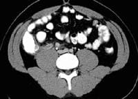 CT scan reveals an enlarged appendix with thickened walls, which do not fill with colonic contrast agent, lying adjacent to the right psoas muscle. The use of CT has dramatically increased since the introduction of multidetector CT (MDCT) scanners. A large, single center study found that MDCT has a high rate of sensitivity and specificity (98.5% and 98%, respectively) for diagnosing acute appendicitis.[37]
Concerns have grown over the possible adverse effects on patients from exposure to radiation from CT scanning. Ultrasonography may offer a safer alternative as a primary diagnostic tool for appendicitis, with CT scanning used in those cases in which ultrasonograms are negative or inconclusive
Go to Imaging of Appendicitis for more information on this topic.
Studies have found a decrease in negative laparotomy rate and appendiceal perforation rate when pelvic CT imaging was used in selected patients with suspected appendicitis.[33, 34, 35, 36] An enlarged appendix is shown in the CT below.
CT scan reveals an enlarged appendix with thickened walls, which do not fill with colonic contrast agent, lying adjacent to the right psoas muscle. The use of CT has dramatically increased since the introduction of multidetector CT (MDCT) scanners. A large, single center study found that MDCT has a high rate of sensitivity and specificity (98.5% and 98%, respectively) for diagnosing acute appendicitis.[37] Concerns have grown over the possible adverse effects on patients from exposure to radiation from CT scanning. Ultrasonography may offer a safer alternative as a primary diagnostic tool for appendicitis, with CT scanning used in those cases in which ultrasonograms are negative or inconclusive
Go to Imaging of Appendicitis for more information on this topic.
Ultrasonography
Because of concerns about patient exposure to radiation during CT scans, ultrasonography has been suggested as a safer primary diagnostic modality for appendicitis, with CT scanning used secondarily when ultrasonograms are negative or inconclusive.[38, 39, 40]
In pediatric patients, the ACEP 2010 clinical policy update recommends using ultrasonography for confirmation, but not exclusion, of acute appendicitis. To definitively exclude acute appendicitis, CT is recommended.[6, 7]
A healthy appendix usually cannot be viewed with ultrasonography. When appendicitis occurs, the ultrasonogram typically demonstrates a noncompressible tubular structure of 7-9 mm in diameter (see the images below).
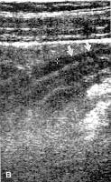 Sagittal graded compression transabdominal sonogram shows an acutely inflamed appendix. The tubular structure is noncompressible, lacks peristalsis, and measures greater than 6 mm in diameter. A thin rim of periappendiceal fluid is present.
 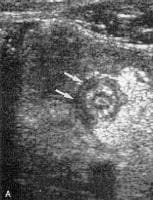 Transverse graded compression transabdominal sonogram of an acutely inflamed appendix. Note the targetlike appearance due to thickened wall and surrounding loculated fluid collection. Vaginal ultrasonography alone or in combination with transabdominal scan may be useful to determine the diagnosis in women of childbearing age. One study of 22 pregnant women in the first and second trimesters showed that graded compression ultrasonography had a sensitivity of 66% and specificity of 95%.[41]
Go to Imaging of Appendicitis for more information on this topic.
In pediatric patients, the ACEP 2010 clinical policy update recommends using ultrasonography for confirmation, but not exclusion, of acute appendicitis. To definitively exclude acute appendicitis, CT is recommended.[6, 7]
A healthy appendix usually cannot be viewed with ultrasonography. When appendicitis occurs, the ultrasonogram typically demonstrates a noncompressible tubular structure of 7-9 mm in diameter (see the images below).
Sagittal graded compression transabdominal sonogram shows an acutely inflamed appendix. The tubular structure is noncompressible, lacks peristalsis, and measures greater than 6 mm in diameter. A thin rim of periappendiceal fluid is present. Transverse graded compression transabdominal sonogram of an acutely inflamed appendix. Note the targetlike appearance due to thickened wall and surrounding loculated fluid collection. Vaginal ultrasonography alone or in combination with transabdominal scan may be useful to determine the diagnosis in women of childbearing age. One study of 22 pregnant women in the first and second trimesters showed that graded compression ultrasonography had a sensitivity of 66% and specificity of 95%.[41] Go to Imaging of Appendicitis for more information on this topic.
Abdominal Radiography
The kidneys-ureters-bladder (KUB) radiographic view is typically used to visualize an appendicolith in a patient with symptoms consistent with appendicitis (see the following image). This finding is highly suggestive of appendicitis, but appendicoliths also occur in fewer than 10% of cases. The consensus in the literature is that plain radiographs are insensitive, nonspecific, and not cost-effective.
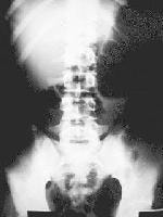 Kidneys-ureters-bladder (KUB) radiograph shows an appendicolith in the right lower quadrant. An appendicolith is seen in fewer than 10% of patients with appendicitis, but, when present, it is essentially pathognomonic. Go to Imaging of Appendicitis for more information on this topic.
Kidneys-ureters-bladder (KUB) radiograph shows an appendicolith in the right lower quadrant. An appendicolith is seen in fewer than 10% of patients with appendicitis, but, when present, it is essentially pathognomonic. Go to Imaging of Appendicitis for more information on this topic.Barium Enema Study
In the past, barium enema examination was used to diagnose appendicitis; in the era of ultrasonography and CT scanning, barium enema study has essentially no role in the diagnosis of acute appendicitis.
A single-contrast study can be performed on an unprepared bowel. Absent or incomplete filling of the appendix coupled with pressure effect or spasm in the cecum suggests appendicitis. The typical radiologic sign of appendicitis is the "reverse 3," which typically manifests as an indentation of the cecum. However, the appendix cannot be visualized in 50% of healthy individuals; therefore, barium enema lacks reliability.
Go to Imaging of Appendicitis for more information on this topic.
Radionuclide Scanning
Whole blood is withdrawn for radionuclide scanning. Neutrophils and macrophages are labeled with technetium Tc 99m (99m Tc) albumin and administered intravenously. Then, images of the abdomen and pelvis are obtained serially over 4 hours. Localized uptake of tracer in the RLQ suggests appendiceal inflammation; this is shown in the images below.
Technetium-99m radionuclide scan of the abdomen shows focal uptake of labeled WBCs in the right lower quadrant consistent with acute appendicitis. Go to Imaging of Appendicitis for more information on this topic.
Technetium-99m radionuclide scan of the abdomen shows focal uptake of labeled WBCs in the right lower quadrant consistent with acute appendicitis. Go to Imaging of Appendicitis for more information on this topic. MRI
Magnetic resonance imaging (MRI) plays a relatively limited role in the evaluation of appendicitis because of its high cost, long scan times, and limited availability. However, the lack of ionizing radiation makes it an attractive modality in pregnant patients. In fact, Cobben et al showed that MRI is far superior to transabdominal ultrasonography in evaluating pregnant patients with suspected appendicitis.[42]
Nonetheless, when evaluating pregnant patients with suspected appendicitis, graded compression ultrasonography should be the imaging test of choice. If ultrasonography demonstrates an inflamed appendix, the patient should undergo appendectomy. If graded compression ultrasonography is nondiagnostic, the patient should undergo MRI of the abdomen and pelvis.
Go to Imaging of Appendicitis for more information on this topic.
Gross and Microscopic Evaluation
In the early stages of appendicitis, the appendix grossly appears edematous with dilation of the serosal vessels. Microscopy demonstrates neutrophil infiltrate of the mucosal and muscularis layers extending into the lumen. As time passes, the appendiceal wall grossly appears thickened, the lumen appears dilated, and a serosal exudate (fibrinous or fibrinopurulent) may be observed as granular roughening. At this stage, mucosal necrosis may be observed microscopically.
At the later stages of appendicitis, the appendix grossly shows marked signs of mucosal necrosis extending into the external layers of the appendiceal wall that can become gangrenous. Sometimes, the appendix may be found in a collection of pus. At this stage of appendicitis, microscopy may demonstrate multiple microabscesses of the appendiceal wall and severe necrosis of all layers.
At the later stages of appendicitis, the appendix grossly shows marked signs of mucosal necrosis extending into the external layers of the appendiceal wall that can become gangrenous. Sometimes, the appendix may be found in a collection of pus. At this stage of appendicitis, microscopy may demonstrate multiple microabscesses of the appendiceal wall and severe necrosis of all layers.
Approach Considerations
Appendectomy remains the only curative treatment of appendicitis, but management of patients with an appendiceal mass can usually be divided into the following 3 treatment categories:
Go to Pediatric Appendicitis for more information on this topic.
- Patients with a phlegmon or a small abscess: After intravenous (IV) antibiotic therapy, an interval appendectomy can be performed 4-6 weeks later.
- Patients with a larger well-defined abscess: After percutaneous drainage with IV antibiotics is performed, the patient can be discharged with the catheter in place. Interval appendectomy can be performed after the fistula is closed.
- Patients with a multicompartmental abscess: These patients require early surgical drainage.
Go to Pediatric Appendicitis for more information on this topic.
Emergency Department Care
The emergency department (ED) clinician must evaluate the larger group of patients who present to the ED with abdominal pain of all etiologies with the goal of approaching 100% sensitivity for the diagnosis in a time-, cost-, and consultation-efficient manner.
Establish IV access and administer aggressive crystalloid therapy to patients with clinical signs of dehydration or septicemia. Patients with suspected appendicitis should not receive anything by mouth.
Administer parenteral analgesic and antiemetic as needed for patient comfort. The administration of analgesics to patients with acute undifferentiated abdominal pain has historically been discouraged and criticized because of concerns that they render the physical findings less reliable. However, at least 8 randomized controlled studies have demonstrated that administering opioid analgesic medications to adult and pediatric patients with acute undifferentiated abdominal pain is safe; no study has shown that analgesics adversely affect the accuracy of physical examination.[43]
Consider ectopic pregnancy in women of childbearing age, and obtain a qualitative beta–human chorionic gonadotropin (beta-hCG) measurement in all cases.
Administer intravenous antibiotics to those with signs of septicemia and to those who are to proceed to laparotomy.
Nonsurgical Treatment
Nonsurgical treatment may be useful when appendectomy is not accessible or when it is temporarily a high-risk procedure. Anecdotal reports describe the success of IV antibiotics in treating acute appendicitis in patients without access to surgical intervention (eg, submariners, individuals on ships at sea).
In a prospective study of 20 patients with ultrasonography-proven appendicitis, symptoms resolved in 95% of patients receiving antibiotics alone, but 37% of these patients had recurrent appendicitis within 14 months.[44]
In a prospective study of 20 patients with ultrasonography-proven appendicitis, symptoms resolved in 95% of patients receiving antibiotics alone, but 37% of these patients had recurrent appendicitis within 14 months.[44]
Preoperative Antibiotics
Preoperative antibiotics have demonstrated efficacy in decreasing postoperative wound infection rates in numerous prospective controlled studies, and they should be administered in conjunction with the surgical consultant. Broad-spectrum gram-negative and anaerobic coverage is indicated (see Medications).
Penicillin-allergic patients should avoid beta-lactamase type antibiotics and cephalosporins. Carbapenems are a good option in these patients.
Pregnant patients should receive pregnancy category A or B antibiotics.
Penicillin-allergic patients should avoid beta-lactamase type antibiotics and cephalosporins. Carbapenems are a good option in these patients.
Pregnant patients should receive pregnancy category A or B antibiotics.
Urgent Versus Emergent Appendectomy
A retrospective study suggested that the risk of appendiceal rupture is minimal in patients with less than 24-36 hours of untreated symptoms,[45] and another retrospective study suggested that appendectomy within 12-24 hours of presentation is not associated with an increase in hospital length of stay, operative time, advanced stages of appendicitis, or complications compared with appendectomy performed within 12 hours of presentation.[46]
Additional studies are needed to demonstrate whether initiation of antibiotic therapy followed by urgent appendectomy is as effective as emergent appendectomy for patients with unperforated appendicitis.
Go to Appendectomy and Pediatric Appendectomy for more information on these topics.
Additional studies are needed to demonstrate whether initiation of antibiotic therapy followed by urgent appendectomy is as effective as emergent appendectomy for patients with unperforated appendicitis.
Go to Appendectomy and Pediatric Appendectomy for more information on these topics.
Emergent Versus Interval Surgery for Perforated Appendicitis
Historically, immediate (emergent) appendectomy was recommended for all patients with appendicitis, whether perforated or unperforated. More recent clinical experience suggests that patients with perforated appendicitis with mild symptoms and localized abscess or phlegmon on abdominopelvic computed tomography (CT) scans can be initially treated with IV antibiotics and percutaneous or transrectal drainage of any localized abscess. If the patient's symptoms, WBC count, and fever satisfactorily resolve, therapy can be changed to oral antibiotics and the patient can be discharged home. Then, delayed (interval) appendectomy can be performed 4-8 weeks later.
The above approach is successful in the vast majority of patients with perforated appendicitis and localized symptoms. Some have suggested that interval appendectomy is not necessary, unless the patient presents with recurrent symptoms. Further studies are needed to clarify not only whether routine interval appendectomy is indicated but also to identify the optimal treatment strategy in patients with perforated appendicitis
Go to Appendectomy and Pediatric Appendectomy for more information on these topics.
Laparoscopic Appendectomy
Initially performed in 1987, laparoscopic appendectomy has been performed in thousands of patients and is successful in 90-94% of attempts. It has also been demonstrated that laparoscopic appendectomy is successful in approximately 90% of cases of perforated appendicitis. However, this procedure is contraindicated in patients with significant intra-abdominal adhesions.
According to the 2010 Society of American Gastrointestinal and Endoscopic Surgeons (SAGES) guideline, the indications for laparoscopic appendectomy are identical to those for open appendectomy.[47]
The 2010 SAGES guideline lists the following conditions as suitable for laparoscopic appendectomy:[47]
Diagnostic laparoscopy may be useful in selected cases (eg, infants, elderly patients, female patients) to confirm the diagnosis of appendicitis. This procedure has been suggested for pregnant patients in the first trimester with suspected appendicitis. If findings are positive, such procedures should be followed by definitive surgical treatment at the time of laparoscopy. Although negative appendectomy does not appear to adversely affect maternal or fetal health, diagnostic delay with perforation does increase fetal and maternal morbidity. Therefore, aggressive evaluation of the appendix is warranted in this group.
According to the SAGES guideline, if findings are negative (normal appendix) on laparoscopic approach, removal should be considered based on the patient’s clinical situation.[47]
Advantages of laparoscopic appendectomy include increased cosmetic satisfaction and a decrease in the postoperative wound-infection rate. Some studies show that laparoscopic appendectomy shortens the hospital stay and convalescent period compared with open appendectomy.
Disadvantages of laparoscopic appendectomy are increased cost and an operating time approximately 20 minutes longer than that of an open appendectomy; however, the latter may resolve with increasing experience with laparoscopic technique. The SAGES guideline recommends practicing a consistent operative method to reduce cost, operating time, and complications.[47]
Go to Appendectomy and Pediatric Appendectomy for more information on these topics.
According to the 2010 Society of American Gastrointestinal and Endoscopic Surgeons (SAGES) guideline, the indications for laparoscopic appendectomy are identical to those for open appendectomy.[47]
The 2010 SAGES guideline lists the following conditions as suitable for laparoscopic appendectomy:[47]
- Uncomplicated appendicitis
- Appendicitis in pediatric patients
- Suspected appendicitis in pregnant women
- Perforated appendicitis
- Appendicitis in elderly patients
- Appendicitis in obese patients
Diagnostic laparoscopy may be useful in selected cases (eg, infants, elderly patients, female patients) to confirm the diagnosis of appendicitis. This procedure has been suggested for pregnant patients in the first trimester with suspected appendicitis. If findings are positive, such procedures should be followed by definitive surgical treatment at the time of laparoscopy. Although negative appendectomy does not appear to adversely affect maternal or fetal health, diagnostic delay with perforation does increase fetal and maternal morbidity. Therefore, aggressive evaluation of the appendix is warranted in this group.
According to the SAGES guideline, if findings are negative (normal appendix) on laparoscopic approach, removal should be considered based on the patient’s clinical situation.[47]
Advantages of laparoscopic appendectomy include increased cosmetic satisfaction and a decrease in the postoperative wound-infection rate. Some studies show that laparoscopic appendectomy shortens the hospital stay and convalescent period compared with open appendectomy.
Disadvantages of laparoscopic appendectomy are increased cost and an operating time approximately 20 minutes longer than that of an open appendectomy; however, the latter may resolve with increasing experience with laparoscopic technique. The SAGES guideline recommends practicing a consistent operative method to reduce cost, operating time, and complications.[47]
Go to Appendectomy and Pediatric Appendectomy for more information on these topics.
Complications
Complications of appendicitis may include wound infection, dehiscence, bowel obstruction, abdominal/pelvic abscess, and, rarely, death. Stump appendicitis also occurs rarely; however, at least 36 reported cases of appendicitis in the surgical stump after previous appendectomy exist.[48]
Consultations
In cases of suspected appendicitis, consult a general surgeon. The surgeon's goals are to evaluate a relatively small population of patients referred for suspected appendicitis and to minimize the negative appendectomy rate without increasing the incidence of perforation.
Không có nhận xét nào:
Đăng nhận xét